Mycoplasma
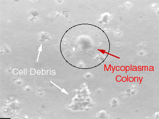
Mycoplasma “colony” on a special formula agar plate @ 100x magnification.
Mycoplama does not grow as colonies in cell cultures and cannot be detected by light microscopy.

Description: Mycoplasmas (PPLO) are small (0.2 – 0.3 micron) wall-less bacteria that can grow to very high concentrations in mammalian cell cultures, typically 107 – 108 organisms/ml, while remaining unobservable by regular light microscopy. Although late stage mycoplasma contamination can cause cell culture medium to become acidic, usually there are no overt signs that cultures are contaminated other than described below.
Characteristics in mammalian cell cultures: In early stages of contamination, mycoplasmas do not typically cause pH changes in the medium nor do they have significant toxic effects on mammalian cells. Mycoplasmas grow mostly associated with the mammalian cell membranes. In many cases there are no signs of mycoplasma contamination, however, mycoplasma can cause changes in cell growth characteristics, inhibition of cell metabolism, disruption of nucleic acid synthesis, chromosomal abberations, changes in cell membrane antigenicity, and can alter transfection rates and virus susceptability. The only way to confirm mycoplasma contamination is by routine testing using special techniques. The nature of the tests available leads the TCF to recommend using two types of tests for a confirming result while using only one type of test should be considered a presumptive result.
Typical routes of infection in cultures: In most cases mycoplasma contamination occurs through cross-contamination from untested infected cells to other cell lines. The route is typically via airborne microscopic aerosolization during pipetting or some other transfer of medium and/or cells during routine handling when more than one cell line is under the hood at a time or when the same bottle of medium is used for more than one cell line. The best prevention is good aseptic technique in conjunction with routine testing. Always try to work “clean-to-dirty” in order of handling cultures during a work day or week. That is, handle confirmed uncontaminated cells first, unknown or untested cells next, and lastly cells that are suspected or known to be contaminated but must be retained in the lab for special reasons.
Antibiotics: Most routine antibiotics used in cell culture are not effective against mycoplasma. Gentamicin sulfate, kanamycin sulfate, and tylosin tartrate are somewhat effective but may only be inhibitive rather than mycoplasmacidal. A treatment product from Roche Molecular Biochemicals, B-M Cyclin, and the antibiotic ciprofloxacin are two of the most effective treatments for attempting to cure mycoplasma infections but generally have success rates under 80% to 85%. A very useful list of antibiotics, the organisms they are effective against, and recommended working concentrations compiled by the Sigma-Aldrich Company can be found as a pdf document (129Kb, 3 pages) by clicking on the following link. The file will be downloaded and can be opened with Adobe Acrobat Reader. Antibiotic List
