
UNC Lineberger researchers are studying a new form of ultrasound that can image the abnormal blood vessels feeding tumors. In a new study, they used this ultrasound technique to show that the abnormal blood vessels feeding cancer tumors extend beyond the tumor borders.
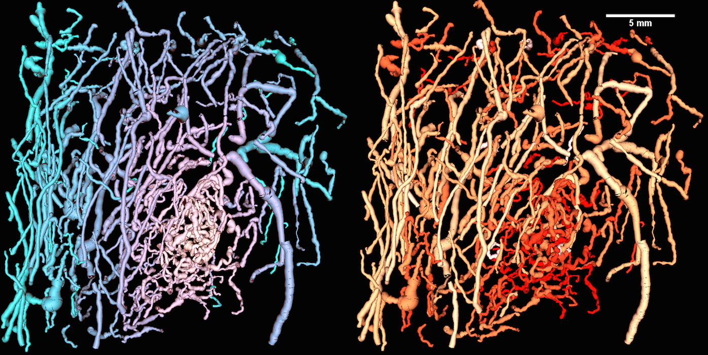
Cancer tumors need nutrients to support their uncontrolled growth, so they stimulate the body to produce new blood vessels. And while normal vessels grow in a tree-like pattern, researchers have found that the blood vessels supporting cancer tumors are abnormally “bendy” and dense.
UNC Lineberger Comprehensive Cancer Center researchers are using the unusual bendiness, or “tortuosity,” of blood vessels supplying tumors to study a potential new cancer imaging technique. They have developed a high-resolution, non-invasive ultrasound technology, which uses sound to create images.
Called “acoustic angiography,” their technology involves bouncing sound waves off microbubbles that are flowing through the blood vessels. When they send sound waves into the body, the waves hit the microbubbles, the bubbles vibrate, and send sound waves back at a different frequency than the rest of the surrounding tissue. The researchers can capture those sound waves distinctly, and translate them into images.
“It is pretty well believed that angiogenesis, the development of new blood vessels, is associated with malignant cancers,” said Paul Dayton, PhD, a UNC Lineberger member and a professor at the UNC-Chapel Hill and North Carolina State University Joint Department of Biomedical Engineering. “However, to-date, there have been few non-invasive imaging methods that are capable of imaging microvasculature with sufficient resolution and contrast to enable analysis of the vessel morphology. We now have a tool that can do this.”
In a new study, the researchers used this ultrasound technique to show that the abnormal blood vessels feeding cancer tumors extend beyond the tumor borders. The researcher say the findings, which were published in September in the Institute of Electrical and Electronics Engineers (IEEE) journal Transactions on Biomedical Engineering, have potential clinical significance.
“Our data suggests that the ‘abnormality’ that we can detect associated with cancer has a larger physical area than the solid tumor itself, which means it might be easier to detect cancers which would otherwise be below our resolution – or that are too small to detect — because the ‘fingerprint’ of abnormal vasculature is larger,” said Dayton, who was the study’s senior author.
The finding is the latest for the researchers in a more than five-year effort. They reported their first findings from a preclinical study that used the technology in the journal Radiology in 2012. And in April of this year, they reported in the journal Ultrasound in Medicine and Biology that they found a quantitatively higher tortuosity and density of blood vessels associated with spontaneously developing breast cancer in a preclinical study.
And now, they are working on getting a clinical trial off the ground to test the technology in human patients with suspicious breast lesions at the N.C. Cancer Hospital. They are hoping to find out whether they can use acoustic angiography to reduce the numbers of false-positive tests for breast cancer and discriminate lethal cancers from non-lethal disease.
In addition to Dayton, other authors of the paper published in IEEE include: Sneha R. Rao, of the Joint Department of Biomedical Engineering; and Sarah E. Shelton, a research assistant in the Joint Department of Biomedical Engineering.
Funding was provided by the National Institutes of Health through grant numbers 1R01CA170665 and 1R43CA165621, P30CA16086, and T32HL069768.