Technologies
The Pathology Services Core may offer additional technologies not listed. For more information, contact us by telephone at 919-445-4179 or e-mail gdelacruz@med.unc.edu.
Spatial Profiling
GeoMx Digital Spatial Profiler (NanoString)
The GeoMx Digital Spatial Profiler provides spatial context to tissue by combining high plex gene expression profiling with the spatial context of immunohistochemistry. This system has the capability to quantify up to 96 proteins and over 1000 RNA targets with spatial context, process 5 slides per day, and preserve precious samples with non-destructive processing.
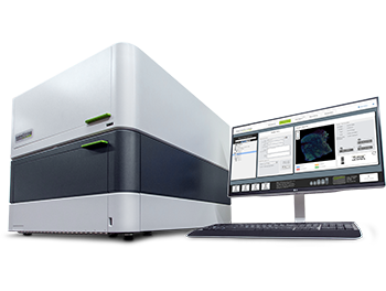
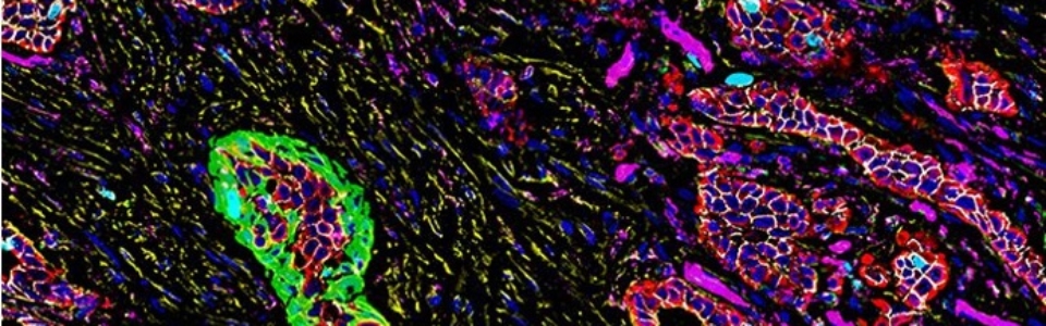
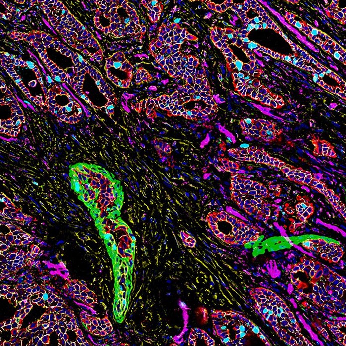
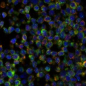
Phenocycler-Fusion (Akoya)
The PhenoCycler™-Fusion system is the fastest spatial biology solution that enables ultrahigh-plex spatial phenotyping of whole slides at single-cell resolution by integrating automated fluidics and iterative imaging.
Researchers are thus able to visualize around 50+ protein targets on one slide. RNA targeting protocols are in the making and will be available
soon.
Visium Manual or CytAssist (10X Genomics)
isium Spatial Gene Expression is a next-generation molecular profiling solution for classifying tissue based on total mRNA. Map the whole transcriptome with morphological context in FFPE or fresh frozen tissues to discover novel insights into normal development, disease pathology, and clinical translational research.
The Visium CytAssist is a compact instrument designed to simplify the Visium workflow. Maximize precious samples—analyze RNA & histology on the same tissue section.
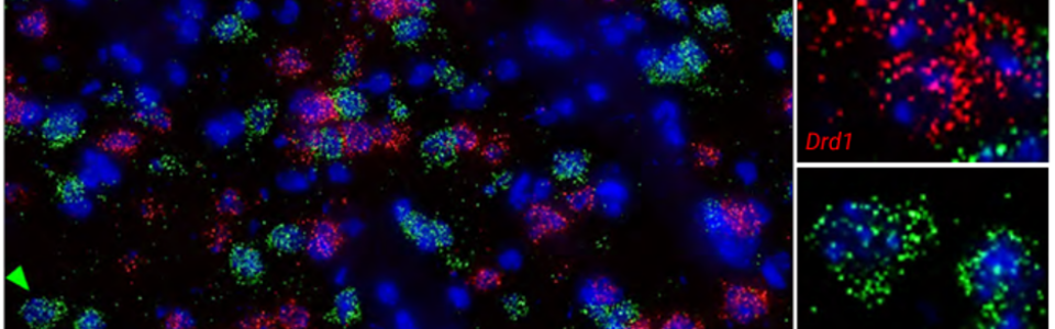
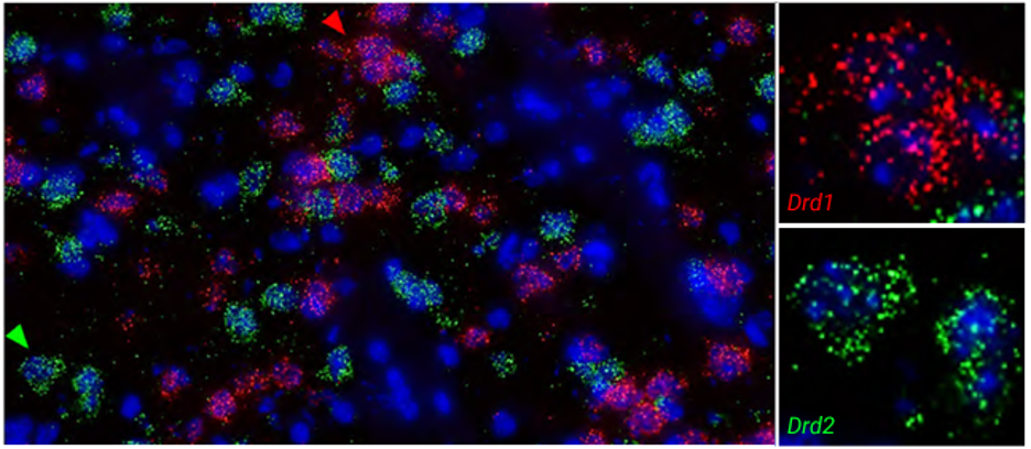
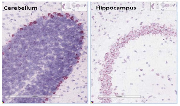
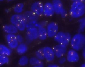
RNAScope (ACD/Biotechne)
The RNAscope assay, a novel multiplex nucleic acid ISH technology with a patented probe design, amplifies target-specific signals but not background noise, delivering clear and actionable results with high sensitivity and specificity. Its signature “double Z” probe design allows for the spatial interrogation of mRNA and long non‑coding RNA (lncRNA) targets longer than 300 nucleotides in any tissue and any species.
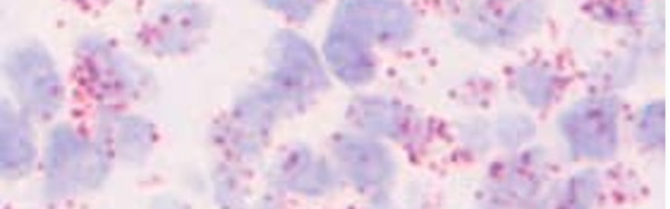
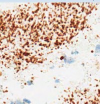
BaseScope (ACD/Biotechne)
BaseScope assay has been developed to spatially map RNA molecules that have previously been unable to be detected in the tissue context. This powerful ISH assay enables the specific detection of exon junctions, splice variants, highly homologous sequences, short targets (~50 to 300 nucleotides), and point mutations, all with single molecule detection sensitivity at single cell resolution in a broad range of tissues, samples and species.
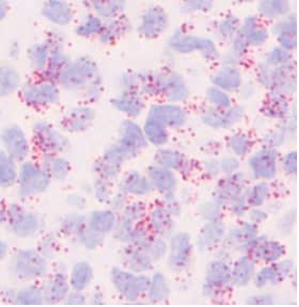
miRNAScope (ACD/Biotechne)
The miRNAscope™ Assay is a highly sensitive and specific in situ hybridization assay that allows for the visualization of ASO, miRNA, siRNA and other nucleic acid targets between 17-50 nucleotides expression in intact tissues or cultured cells with single-cell resolution, preserving spatial and morphological context.
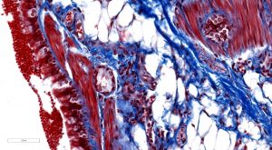
Staining
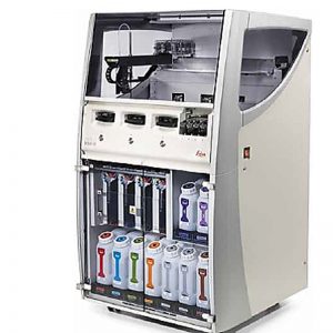
Bond III & Bond Rx Autostainers (Leica Biosystems)
The Bond II and Bond Rx autostainer systems have the capacity to run 30 slides in a given run, with run times varying between 15 minutes to 6 hours. Triplex staining is also possible on this equipment.

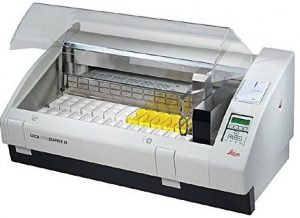
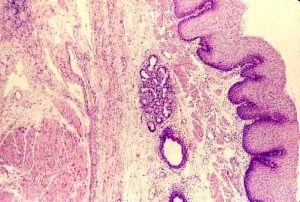
XL Autostainer (Leica Biosystems)
The Leica Biosystems autostainer allows for automated and programmable deparaffinization of slides and hematoxylin and eosin (H&E) staining.
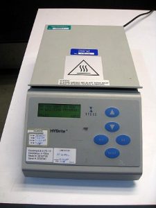
HYBrite Slide Stainer (Vysis)
The HYBrite Slide Stainer can be programmed to hybridize, denature, and humidify up to 12 slide per run.
Discovery Ultra Ventana (Roche)

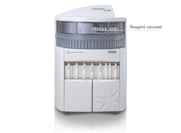
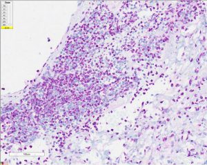
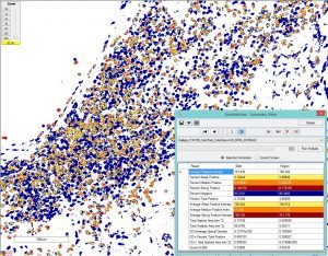
Image Analysis
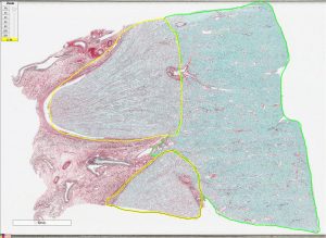
Image Analysis with Aperio Toolbox/GENIE, Definiens Tissue Studio, and Visiopharm
PSC has contributed to an average of 29 publications per year.
Slide Scanning
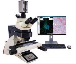
Versa 200 (Leica Biosystems)
Brightfield and fluorescence tiling scanner with a 200 slide capacity. Can scan at 1.25X, 5X, 10X, 20X, 40X, and 63X oil; DAPI/Hoechst, FITC/Alexa 488, Cy3/Alexa 560, Spectrum Orange, Texas Red/Alexa 594, Cy5/Alexa 647, and Cy7/Alexa 750 filter cubes. Standard file format is Leica SCN.
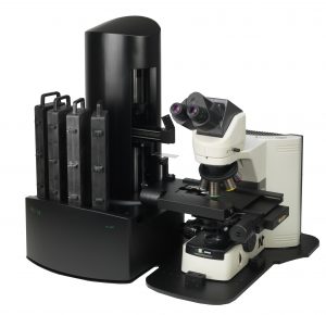
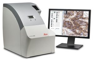
ScanScope AT2 (Leica Biosystems)
Brightfield line scanner with a 400 slide capacity. Can scan at 20X or 40X, with a standard file format of Aperio SVS.
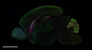
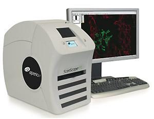
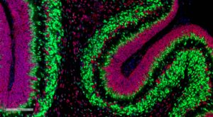
ScanScope FL (Leica Biosystems)
Fluorescence line scanner with a 5 slide capacity. Can scan at 20X or 40X; DAPI/Hoechst, Spectrum Aqua, FITC/Alexa 488, Cy3/Alexa 560, and Cy5/Alexa 647 filter cubes. Standard file format is AFI.
Histology
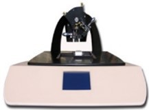
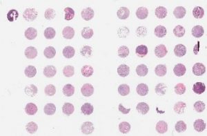
Semi-Automated Tissue Arrayer
This system can make up to four TMA blocks simultaneously, using human or animal tissues, cell lines, or tissue reconstructs, with up to 300 cores per TMA block possible. The tissue core diameters range over 0.6, 1.0, and 2.0mm.
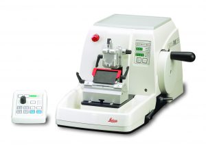
Microtomes (Leica Biosystems)
The Leica Biosystems Microtome holds the capability to consistently cut paraffin tissue sections in a 4-20 micrometer thick range.
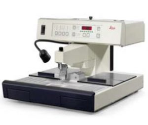
Tissue Embedding Systems (Leica Biosystems)
The Leica Tissue Embedding station uses paraffin wax to create Formalin-Fixed Paraffin-Embedded (FFPE) tissue blocks. Additional features include manual control, multiple mold sizes, and paraffin block cooling plate.

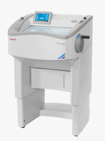
Epredia CryoStar NX70
Labeling Systems
Slide and Tube Label Printers (Brady & CILS)
The Brady/CILS slide and tube label systems create solvent resistant labels as well as temporary labels to be used in the scanning of diagnostic slides. These systems are capable of printing barcodes that are readable by slide scanners. Different label materials are available upon request.