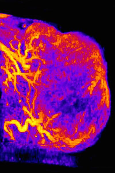
The research, led by Andrew C. Dudley, has implications for developing cancer drugs that target blood vessels that feed tumors.

CHAPEL HILL, NC – UNC School of Medicine researchers have pinpointed a set of intriguing characteristics in a previously unknown subpopulation of melanoma cancer cells in blood vessels of tumors. These cells, which mimic non-cancerous endothelial cells that normally populate blood vessels in tumors, could provide researchers with another target for cancer therapies.
The research, published today in the journal Nature Communications, provides evidence for how these particular melanoma cells help tumors resist drugs designed to block blood vessel formation.
“For a long time the hope has been that anti-angiogenic therapies would starve tumors of the nutrients they need to thrive, but these drugs haven’t worked as well as we all had hoped,” said Andrew C. Dudley, PhD, assistant professor in the Department of Cell Biology and Physiology, member of the UNC Lineberger Comprehensive Cancer Center, and senior author of the paper. “There are likely several reasons why these drugs haven’t been effective; our research suggests that these previously uncharacterized cells could be one of the reasons.”
Most of the drugs developed to disrupt tumor blood vessels target a protein called vascular endothelial growth factor, or VEGF, which is part of a major signaling pathway in the noncancerous endothelial cells that typically line blood vessels in tumors. But other research has suggested that tumors are able to resist anti-angiogenic therapies – particularly those targeting VEGF – through a variety of complex mechanisms. In one set of experiments, Dudley and graduate student James Dunleavey, used a known anti-angiogenic drug which blocks VEGF and found that this new subpopulation of melanoma cells was more prevalent in drug-resistant tumors in mouse tumor models. Moreover, tumors composed entirely by this new subpopulation in mouse models did not respond at all to anti-VEGF therapy.
This research further elucidates the complex nature of human tumors, which are not comprised solely of the same type of cancer cell but instead feature a mixed population of different cell types with different functions.
To make these discoveries, Dunleavey first isolated what he thought were noncancerous endothelial cells from melanoma tumors. Then he conducted a genetic analysis to reveal that these cells did not express most of the well-known biomarkers common to endothelial cells. For instance, the cells didn’t express VEGF receptors. This could explain why the anti-VEGF therapy was ineffective at blocking tumor growth.
“These cells looked very different from normal endothelial cells in cultures,” said Dudley, who is also a member of the UNC McAllister Heart Institute. “We didn’t know what these cells were. For a while, we kind of scratched our heads until Jim conducted more experiments over the course of a year to find that these cells had several markers similar to melanoma cells.” Further analysis revealed that these cells were in fact a new kind of melanoma cell, one that expressed a particular protein called PECAM1, which is very important to the function of endothelial cells.
PECAM1 is an adhesion molecule. Scientists have known that endothelial cells use it to stick together to form blood vessels. When Dudley’s team looked at blood vessels in tumors made of PECAM1-positive tumors, they found melanoma cells alongside noncancerous endothelial cells on the inside of vessels. “We don’t think these new melanoma cells are just passively filling in gaps between endothelial cells in the tumor vasculature,” Dudley said. “How they come to form blood-filled channels seems to be an active process that involves PECAM1.”
Dudley and Dunleavey then teamed up with other scientists, including UNC’s Paul Dayton, PhD, a professor in the Department of Biomedical Engineering, member of the UNC Lineberger Comprehensive Cancer Center, and co-author on the Nature Communications paper. Dayton’s lab conducted ultrasound imaging studies showing that PECAM1-positive tumor blood vessels in mice had twice the vascular density of PECAM1-negative vessels. And the blood volume of PECAM1-positive blood vessels was 4 ½ times greater than PECAM1-negative vessels. This showed the researchers that these newly discovered PECAM1-positive melanoma cells had a real effect on the function of tumor blood vessels.
Dunleavey added, “We think these cells help allow tumor cells to interact in specific ways with bona fide endothelial cells.” And this interaction – plus the lack of VEGF responsiveness – could help blood vessels and blood-filled channels resist therapies designed to destroy them.
Dunleavey and Dudley said that this discovery is likely just one part of how some tumors manage to circumnavigate therapies designed to attack their blood vessels.
“Anti-angiogenic therapies are typically used in conjunction with another drug,” Dunleavey said. “It could be that we’ll need to develop a combination of anti-angiogenic drugs to attack the endothelial cells that form blood vessels and the tumor cells that might form blood-filled channels in tumors.”
###
Other authors of the Nature Communications paper, include UNC graduate students Lin Xiao, David Irwin, Victoria Brings, and Sarah E. Shelton; UNC undergraduate student Joshua Thompson; lab technician Mi Mi Kim; Janiel M. Shields, PhD; David W. Ollila, MD, professor of surgery at UNC; Rolf A. Brekken, PhD, associate professor at the University of Texas Southwestern Medical Center; and Juan M. Melero-Martin, PhD, assistant professor of surgery at Harvard.
This research was funded by grants from the National Institutes of Health and the University Cancer Research Fund at UNC-Chapel Hill.
